
|
Otorhinolaryngology Otorhinolaryngology |
Vertigo and Tinnitus
Tinnitus is the perception of sound in the head or the ears
Vertigo is defined as perception of movement in the absence of movement. It is caused by asymmetry in the baseline input into the vetibular centres, which causes the vertigo but also nystagmus and vomiting
It is very important to differentiate this symptom from other symptoms that patients may report
Typically patients will describe "dizzines", "light-headedness" etc. These are not specific and must be explored further by the doctor and differentiated into:
- pre-syncope
- syncope
- vertigo
- weakness
- unsteadiness or imbalance
Overview
Differential Diagnosis
Central
- Ischaemia: TIA, stroke, vertebrobasilar insufficiency + migraine
- Neoplastic: Acoustic neuroma (usually presents with unilateral progressive hearing loss)
- MS
Peripheral (the first 4 diagnoses are in order of incidence)
- Benign paroxysmal positional vertigo (BPPV)
- Meniere disease
- Vestibular neuronitis
- Labyrinthitis
- Others: otitis media, sinusitis
Note: vertigo that doesn't start to improve within 48 hours may indicate a central cause
History
- As above, ensure you understand exactly which symptomatic entity the patient is describing i.e. is the patient truly describing vertigo?
- Timing: how long does it last?
- Onset: is it sudden?
- Vertigo association: do certain postures bring it about?
- Presence of tinnitus, hearing loss, otalgia, otorrhoea, aural fullness
- Preceding URTI
- Smoking and other medications or herbal remedies
- Review of systems:
- associated gait problems or ataxia (suggest a neurological cause)
- head trauma
- other medical problems
- other ENT diseases
Examination
- Vital signs including orthostatic BP
- Full ENT exam (particularly for signs of infection, hearing loss etc.)
- Thorough neurological exam is mandatory with specific focus on cranial nerves, gait and cerebellar function
Special tests:
- Dix Hallpike test
- Romberg and tandem gait
- Head thrust
- Caloric testing
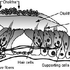
Otolith organs |
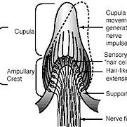
Crista within the semicircular canal |
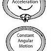
How motion affects the crista |
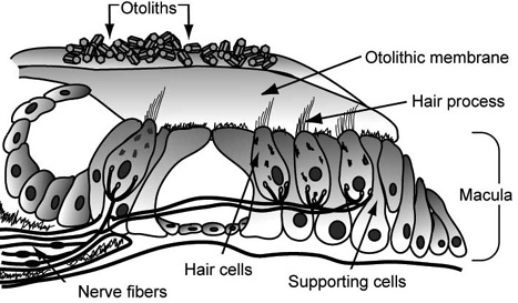
Otolith organs
The saccule and utricle are the otolith organs
The arrangement of the CNVIII nerve fibres, hair cells, otolithic membrane and otoliths is shown in this diagram
The saccule is more sensitive to vertical acceleration and utricle to horizontal acceleration
You may wish to revise this from your special senses notes
The arrangement of the cupulae in the semicircular canals is shown in this diagram.
You may wish to revise this from your special senses notes (3rd year)
When a person's head moves the endolymph moves (or the cupula moves and the fluid remains stationary)
The gelatinous tip of the cupula and the hair cell extensions embedded within it are displaced to one side or the other
When the embedded hair cells bend, they send a signal via the vestibular nerve to the brain where the information is
evaluated and appropriate action is initiated.
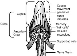
Diagram highlighting the direction of cupula movement with different motions of the head

Investigations
A range of specialist vestibular reflex testing exist
Imaging with MRI is indicated where there is asymmetric hearing loss an (acoustic neuroma may be suspected)
Baseline bloods should be performed e.g. FBC, U+E, Glucose
BPPV
Aetiology
BPPV is defined as vertigo that is elicted by certain head positions. The positions trigger nystagmus.
The cause of this is unknown but is postulated to be due to 2 pathological mechanisms:
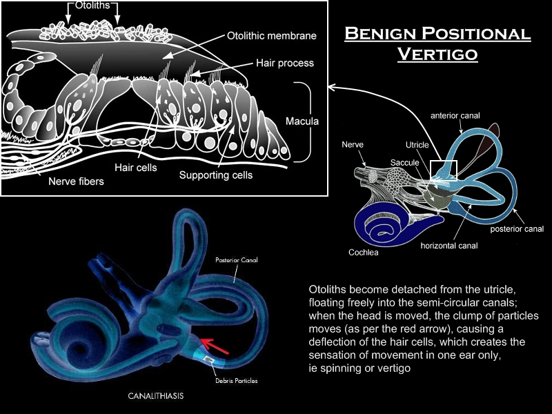
1. Canalithiasis: This is due to otoliths that have become detached from the otolithic organs and are free-floating; exerting a force on the cupula mechanism within the semicircular canals
(analogy is: pebbles in a tyre - when the tyre turns the pebbles tumble down due to gravity; this tumbling triggers the nerves inappropriately)
2. Cupulolithiasis: This is due to deposits of otoliths on the cupula themselves; rendering the cupula more sensitive to gravitational force with certain head positions
(analogy is: heavy object placed on a skinny pole; extra weight makes pole unstable and harder to keep in upright position - pole easily clunks from one side to another and tends to keep tilted position)
History
Onset of vertigo is sudden and severe - lasting ~30 seconds
Typically occur when moving the head in certain positions e.g. from lying to sitting
May be associated with nausea and vomiting
Examination
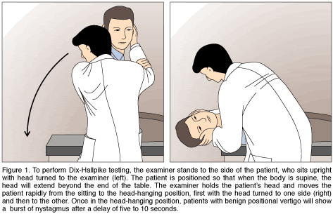
Diagnosis is made by Dix-Hallpike test
Remainder of examination should be normal
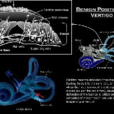
Otoliths in BPPV |
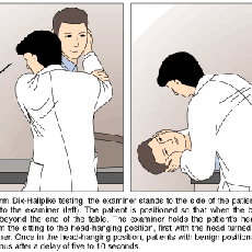
Dix-Hallpike Test |
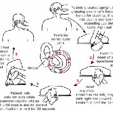
Modified Epley |
Canuloliths
This diagram illustrates the canulolith pathology behind BPPV
Management
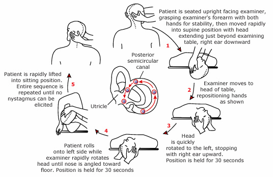
1. Canalith repositioning via Modified Epley Maneuver
2. In the acute setting vestibulosuppresants can be tried for symptomatic relief:
- Promethazine
- Benzodiazepines e.g. lorazepam
- Scopolamine
Surgery reserved for severe intractable cases
Meniere disease
Aetiology
Also known as endolymphatic hydrops (hydrops is another term for oedema)
The cause is thought to be due to impaired reabsorption of the endolymphatic fluid
The precipitent factors is still unknown but postulated to be infectious, immunologic or allergic
History
Meniere disease occurs as attacks lasting for hours. The four symptoms are:
1. Unilateral fluctuating SNHL
2. Vertigo lasting minutes to hours
3. Constant or intermittent tinnitus - typically increasing in intensity during vertiginous attack
4. Aural fullness
The attack is associated with nausea and vomiting - followed by feeling lethargic for a few days
Investigation
Meniere disease is a clinical diagnosis - based on history and normal examination
Audiometry is used to confirm SNHL
The following are performed to help rule out differential diagnoses:
- Syphilis as this can perfectly mimic Meniere disease
- As noted before if hearing loss is asymmetric then MRI needs to be performed
Management
Acutely:
1. Vestibular suppresants - promethazine, benzodiazepines, scopolamine
2. Betahistine
Long term suppression
1. Dietary restriction of salt
2. Thiazide diuretic
3. Betahistine
4. Aminoglycoside injection into middle ear
5. Surgery reserved for severe and intractable cases
Vestibular neuronitis
Aetiology
This is an acute, sustained dysfunction of the peripheral vestibular system causing vertigo, nausea and vomiting
The most likely cause is reactivation of HSV in the vestibular ganglion and nerve - other viruses e.g. adenovirus are also potential pathogens (note: do not confuse this with Herpes Zoster Oticus - which is a Varicella Zoster virus mediated disease)
Epidemiology
Commonly in 4th to 6th decades
History
Commonly have preceding URTI
Sudden and acute vertigo, nausea and vomiting without hearing loss
Lasts for days -> cannot work or do usual activities
Examination
Should have normal hearing and neurological exam (otherwise suspect something more sinister)
Nystagmus will be present with slow phase towards injured ear
Management
1. Vestibular suppressants
2. Corticosteroids for 3 weeks (decreases chances of long term vestibular function loss)
The presence of SNHL indicates that the diagnosis is labyrinthitis. These patients need to be admitted for IV antibiotics
| « Hearing loss | Case » |


