Clinical Practice Case 3
 Case 3
You are examining a 2-year-old patient who was brought to the ED by EMS providers. They report that the child demonstrated increasing respiratory distress. The child was previously well and was found at home with a bottle of lamp oil that he had apparently opened. The mother called EMS immediately when she noted that the toddler was "breathing funny" and appeared in distress. While receiving high-flow oxygen by face mask, his Sp02 during transport and on ED arrival was in the mid to high 80s.
General assessment
You see a tachypneic, anxious toddler who is sitting up and grunting with nasal flaring and signs of increased respiratory effort, specifically intercostal and suprasternal retractions. His colour appears normal. He is alert and watching you anxiously.
3A
What is your initial impression of the child's condition based on your general assessment?
This child's condition is very worrisome because he has significant tachypnea and respiratory distress and he is not responding well to oxygen administration (his pulse oximetry is still in the mid-80s despite oxygen administration).
Does the child need immediate intervention?
He needs rapid interventions to try to improve his oxygenation.
If so, what intervention is indicated?
expiratory pressure (PEEP) to help recruit collapsed alveoli. This may be delivered noninvasively (eg, BiPAP) or through an endotracheal tube during mechanical ventilation.
3B
Grunting is always a worrisome sign. In the setting of respiratory distress, it almost always means the child is trying to keep alveoli and small airways open by increasing end-expiratory pressure. When grunting and hypoxemia are seen together, there is a high likelihood that the child has respiratory failure and will need ventilatory support.
Primary assessment
He is placed on a cardiac monitor and pulse oximeter. His heart rate is 145/min, respirations are 50/min, blood pressure is 115/75 mm Hg, and temperature is 97.8°F (36.5 °C) axillary. His Sp02 is 85% on highflow oxygen. On auscultation you hear moist crackles throughout his lung fields with decreased air entry over his axillary lung fields; there are coarse breath sounds centrally. He has good distal perfusion with readily palpable pulses and brisk capillary refill.
3C
How would you categorize this child's condition?
This child has respiratory distress that is most likely caused by lung tissue (parenchymal) lung injury. This categorization is based on the presence of grunting and hypoxemia that is poorly responsive to oxygen administration. The lack of oxygen responsiveness suggests that the alveoli are not participating in gas exchange because they are collapsed or full of fluid (such as edema fluid or blood). In this case, aspirated lamp oil can injure lung tissue, disrupt surfactant, and stimulate an acute inflammatory reaction (chemical pneumonitis), leading to hypoxemia, pulmonary edema, and decreased lung compliance. The degree of respiratory distress and lack of response to oxygen indicate that the child has respiratory failure.
It is difficult to estimate the degree of hypercarbia in this situationsome children may maintain adequate ventilation even when they have significant hypoxemia. An arterial blood gas is the only way to quantify the adequacy of ventilation. But your clinical exam is often sufficient to recognize when the child is not moving air adequately.
3D
What are your initial decisions and actions?
The lack of response to high-flow oxygen indicates that this child's oxygenation will improve only with the application of positive end-expiratory pressure (PEEP) to help recruit collapsed alveoli. This may be delivered noninvasively (eg, BiPAP) or through an endotracheal tube during mechanical ventilation.
3E
What conditions are associated with the signs and symptoms seen in this child?
Lung tissue disease is caused by a range of conditions producing alveolar collapse or obstruction. The alveoli are filled with inflammatory exudate in children with infectious pneumonia. They may also be filled with edema fluid and varying degrees of inflammatory cells in children with acute respiratory distress syndrome. Children with left heart failure may have alveoli filled with a transudate (low protein-containing fluid) produced from high pulmonary capillary pressures. Other less common causes include aspiration, such as in this case, and acute alveolar hemorrhage.
In view of his marked distress, poor response to oxygen, and the fact that his symptoms are severe so early after the aspiration event, the child's respiratory condition is likely to worsen. As a result, he will need to be electively intubated.
While the equipment is being gathered for intubation and mechanical ventilation, you obtain a SAMPLE history, learning that he rapidly developed breathing §.ymptoms after being found with the lamp oil bottle. Other than the signs noted in the primary assessment, there are no other relevant findings when examining his skin, abdomen, back or neck. You note that his jaw and mouth appear normal. He has no !!.lIergies. He is not receiving any medications, and his Past medical history is unremarkable. He last ate about 2 hours before this event. You do learn a few more details about the event. The toddler's father was refilling several lamps in the garage and left the bottle of lamp oil on the garage floor. The child has no history of other ingestions or unexplained injuries.
3F
What equipment do you need to gather in preparation for intubation?
In addition to the monitoring that is in place, you will need to gather the following:
- Oxygen supply, equipment for connections to airway adjunct device
- Exhaled CO2 detector device
- Suction equipment (large bore suction catheter or tonsil-tipped suction as well as a catheter that can pass through the endotracheal tube)
- Bag-mask connected to a high-flow oxygen source
- Face mask (correct size)
- Endotracheal tubes, proper size (ie, the estimated size and the size 0.5 mm above and below that size)
- Endotracheal tube stylet
- Laryngoscope blade (correct size), curved or straight, with working bulb
- Laryngoscope handle with connector and battery backup light source (another laryngoscope handle and blade)
- Medications
- Commercial endotracheal tube holder type
- Towel or pad to place under the patient's head (to align the airway)
- NG tube (correct size)
- Oropharyngeal airway (correct size)
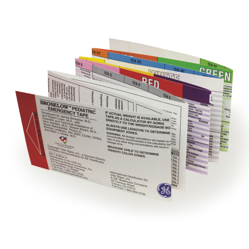
- A length-based resuscitation tape (Broselow Pediatric Emergency Tape) or other reference is helpful to estimate the correct equipment sizes for the child.

Does the likelihood of lung tissue disease change your thoughts about the intubation equipment needed?
When selecting an endotracheal tube for this child, you should consider that he will need increased end-expiratory pressures (ie, positive end-expiratory pressure [PEEP]) and relatively high peak inspiratory pressures, so a cuffed tube would be appropriate if available. If a cuffed tube is used, remember that you must monitor the cuff pressure and maintain it <20 cm H20 to avoid airway injury.
To improve oxygenation prior to intubation, a flow-inflating bag with a tight mask seal may be useful since you can easily provide continuous positive airway pressure (CPAP) with this setup. Alternatively, a PEEP valve may be attached to a self-refilling bag. You should anticipate the need for PEEP following intubation.
3G
What is the significance of the time he last ate?
You must assume that this child's stomach is full. Therefore he is at risk for regurgitation and aspiration of stomach contents. In addition, he may have some of the lamp oil still in his stomach, and you want to avoid a second lamp oil aspiration episode during intubation.
To minimize the risk to this child, the provider with the greatest experience in intubation should perform or closely supervise the intubation procedure. You should use a rapid sequence intubation (RSI) protocol. Once the sedative paralytic has taken effect (and the child loses cough and gag reflexes), a team member should maintain cricoid pressure during attempted intubation.
3H
In addition to gathering equipment, what other preintubation activity should you consider?
Successful resuscitation of this child requires good teamwork. It is important that all members of the team know their roles and that the team leader clearly communicate requests. It is a good idea to assign roles before intubation. Clear communication is important so that the team leader knows what medication has been given and when. It is also essential that you assess the patient's airway anatomy. You should know or have confidence that you can provide effective bag-mask ventilation if needed before giving a neuromuscular blocker as part of a rapid sequence intubation approach.
Case Progression
The child is intubated with a 4.5 mm cuffed endotracheal tube. Tube position is confirmed. You provide mechanical ventilation with 100% oxygen and a PEEP of 6 cm H20. A nasogastric tube is inserted to decompress his stomach. The Sp02 initially was 91 %, and following an increase in the PEEP to 10 cm H20, it improved to 100%. You hear equal breath sounds with bilateral moist crackles but improved air entry over the lateral lung fields. Heart rate is 160/min with a blood pressure of 118/78 mm Hg. Pulses remain strong distally with a good capillary refill. The child is not moving because a neuromuscular blocking agent was used to facilitate intubation.
3I
What tertiary assessment studies are appropriate now?
Following intubation you should obtain a chest x-ray to assess the endotracheal tube depth of insertion and to evaluate the extent of the child's lung disease and the degree of lung inflation.
An arterial blood gas is also indicated to objectively assess the adequacy of ventilation and to compare the child's arterial Paco, with his end-tidal CO2 reading if capnography is being used.
A chest x-ray is obtained, but immediately after the x-ray plate is removed from under the child, his Sp02 and heart rate suddenly fall.
3J
What do you think happened?
In this setting consider causes of sudden deterioration in an intubated child. These can be recalled by the DOPE mnemonic (see Endotracheal Intubation on the student CD). On examination, it is clear that the endotracheal tube was displaced from the trachea.
How would you manage this situation?
You remove the tube and team members provide 2-person bag-mask ventilation using a flow-inflating manual resuscitator with a pressure manometer. You then successfully reintubate the trachea and confirm correct placement with clinical examination and detection of exhaled CO2. The child's oxygenation and ventilation improve immediately following reintubation.
Case Conclusion
Following appropriate intervention, tube position is reconfirmed by clinical examination and capnography. An arterial blood gas is obtained with the following results on 100% oxygen: pH 7.32, PCO2 52, P02 95, base deficit - 0.5.
He is transferred to the pediatric intensive care unit with capnography and pulse oximetry monitoring during transport.
 Summing up
This child had acute lung tissue disease secondary to airway and alveolar injury associated with aspiration of lamp oil into the lungs and resulting lung inflammation. The important point, in this case, is that when severe, this type of injury is likely to require more therapy than oxygen administration alone. Effective therapy often requires mechanical ventilation with the use of positive end-expiratory pressure. Good teamwork is essential to successful intubation and optimal patient outcome.


