Screening tests
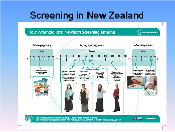
Prenatal screening should be offered to all pregnant women and information for pregnant women and health professionals is available on the National Screening Unit Website which can be accessed at: https://www.nsu.govt.nz/. There are three options for screening for Down's syndrome and other conditions available- two are funded and one is not funded. All women should also have an anatomy scan between 18-20 weeks and follow-up blood tests and diabetes screening between 24-28 weeks.
MSS1 and MSS2
In New Zealand, combined first trimester screening is performed with a blood test (MSS1) and ultrasound for nuchal translucency at 11-13+6 weeks gestation. Together, these tests are combined into an algorithm with the mother’s age, ethnicity, weight, and gestation (along with other variables) to calculate a risk for Downs Syndrome (Trisomy 21), Trisomy 18 and Trisomy 13 respectively. In addition to this, the nuchal translucency may be increased in other conditions such as neural tube defects such as spina bifida. Women with a high risk screening result will be offered a diagnostic test with an amniocentesis. A typical detection rate for Trisomy 21 would be approximately 90% with a false positive rate of 5%.
Alternatively, if women book later in the pregnancy or do not complete both parts of the first trimester screen in the time frame required, they may be offered second trimester screening (MSS2) which is a blood test which also uses a similar algorithm to provide a risk assessment for these trisomies, but with a lower sensitivity and specificity than combined first trimester screening. A typical detection rate for Trisomy 21 would be approximately 75% with a false positive rate of 5%.
In New Zealand, the blood tests for MSS1 OR MSS2 are funded, and there is funding for the nuchal translucency however most ultrasound providers charge the patient a fee for this scan (approximately $40). This may serve as a possible barrier to completed first trimester screening.
NIPT
Non-invasive prenatal testing (NIPT) is a blood test that can be taken from 10 weeks of pregnancy. It is not a publically funded test and is expensive- it can cost up to $1000, although prices are likely to decrease in the future. NIPT is based on the detection of cell free fetal DNA (fragments of fetal DNA) present in maternal blood. A number of techniques are used to evaluate the load of DNA from each chromosome and then predict the likelihood of there being aneuploidy. NIPT is the best available form of prenatal screening for Trisomy 21- it has a 99% detection rate with a false positive rate of less than 1%. However, the cost of this test is a significant barrier to screening for many women.
Diagnostic testing
For women with a positive screen for aneuploidy, diagnostic testing may be offered. There are two options currently available for diagnostic testing- amniocentesis and chorionic villus sampling (CVS). Amniocentesis, which can be done from 14 weeks gestation, is the gold-standard test and involves testing a sample of amniotic fluid. The risk of miscarriage with this procedure is low- 0.4%. CVS can be performed earlier, from 10-11/40 and involves taking a sample of chorionic villi. It is a more difficult procedure, but has a low miscarriage rate- <1%. 1-2% will have placental mosaicism, which means the sample will not be reflective of the fetal karyotype.
18-20 Week Morphology (Anatomy) Ultrasound
The purpose of the morphology ultrasound is to ensure that fetal anatomy is normal and provide referral to a maternal fetal medicine service if fetal anomaly is present. This ultrasound is a screening test for anatomical defects of the fetus, the maternal ovaries and uterus, and the placenta. Ultrasound is very accurate at identifying some anatomical defects, and less accurate in others. Overall, ultrasound can detect approximately 50% of fetal abnormalties in a low risk population when done at 19-20 weeks. Ultrasound is very good at detecting neural tube defects, urinary system defects, and anterior abdominal wall defects, which detection rates of 80-90%. Cardiac, facial, intestinal, and minor skeletal abnormalities are more difficult to detect with ultrasound.
24-28 week blood tests
Other screening tests occur between 24-28 weeks. The full blood count and ferritin are re-tested at that time to screen for the development of anaemia in pregnancy. Detection at this time means that iron supplementation can start and have an effect prior to birth. Repeat testing is performed to check blood group and antibody status to ensure that no new antibodies have developed in the pregnancy. Diabetes testing is also performed at this time. Low risk women have "polycose" testing- this is a 50g glucose load which is given at any time of the day, followed by a blood test at 1 hour after the glucose load. If this is negative and there are no other signs of diabetes in the pregnancy, no other testing is required. If it is positive then a 75g oral glucose tolerance test (OGTT) is performed. High risk women (with risk factors for diabetes or a positive polycose test) have a fasting blood test taken (and they must have fasted for 8 hours prior to the test) and then a 75g glucose load. A second blood test is taken at 2 hours. If this test is negative then no other testing is required, unless symptoms of diabetes develop in the pregnancy. If it is positive then it confirms the diagnosis of gestation diabetes.


