")

Preview
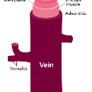
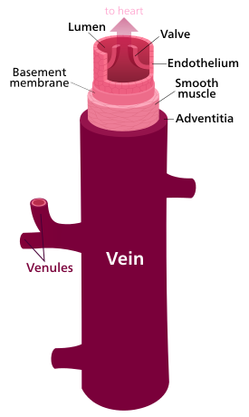
In general, the function of veins is to return deoxygenated blood to the heart, and are tubes that collapse when their lumens are not filled with blood (in hypovolaemic shock).
The thick outermost layer of a vein is made of connective tissue, provides support to the vein and contains capillaries called tunica adventitia or tunica externa.
The middle layer is strands of smooth muscle cells, elastic fibres and connective tissue called tunica media, which are, in general, thin, as veins do not function primarily in a contractile manner:
- Source of pain upon insertion of cannulae
The interior is lined with endothelial cells called tunica intima:
- Sensitive to medications outside the normal pH range of 7.35-7.45 (acidic or basic)
- Sensitive to hypertonic solutions (>275-295 Osmoles/kg)
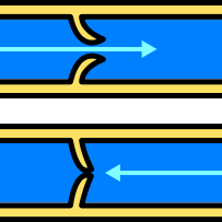
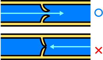
Most veins have one-way flaps called venous valves that prevent blood from flowing back and pooling in the lower extremities due to the effects of gravity. These are infoldings of the tunica intima.
The exact location of the veins is much more variable from person to person than that of arteries.
|
Vein, by Kelvinsong, http://en.wikipedia.org/wiki/File:Vein.svg |
Venous Valve, by ZooFari, http://en.wikipedia.org/wiki/File:Venous_valve.svg |