Respiratory Failure
The term ‘Respiratory System’ not only refers to the lungs but also includes parts of the CNS, peripheral nervous system and musculoskeletal system, all involved in the process of respiration.
The principal function of the respiratory system is to provide gas exchange appropriate to the body’s needs. This involves the uptake of oxygen and removal of carbon dioxide produced by the body’s metabolic process.
‘Respiratory failure’ occurs when the respiratory system cannot meet the body’s gas exchange requirements.
Breathing entails the contraction of respiratory muscles to generate a negative intrathoracic pressure. This entrains environmental air into the lungs. Once inside the lung, oxygen moves across the alveolar membrane into the blood, and carbon dioxide (CO2) is released from the blood into the alveoli. The oxygen is delivered to the body via circulation, and carbon dioxide is excreted during exhalation.
Respiratory failure may be divided into two types:
Oxygenation failure,(Type I) which occurs when the normal oxygen content of the blood cannot be maintained by the lungs’ usual compensatory mechanisms.
Ventilatory failure,(Type II) which refers to inadequate alveolar ventilation resulting in increased carbon dioxide levels and reduced oxygen levels in the body.

Oxygenation Failure
In the normal functioning lung, ventilation—or air delivery to the alveoli—matches the blood flow in the alveolar capillaries.
Hypoxia and respiratory failure essentially occur when the air and blood flow in the lung is unbalanced, or when in some parts of the lung ventilation is greater than blood flow (increase in physiological dead space), and in other parts of the lung blood flow is greater than ventilation. This eventually results in low oxygen levels or hypoxia.
This process is called ‘ventilation/perfusion mismatch’ (V/Q mismatch). The body’s compensatory mechanism in response to V/Q mismatch is to increase ventilation, which only partially corrects blood oxygenation.
Disease processes in the lung that cause hypoxic respiratory failure can be broadly grouped into:
parenchymal diseases
airway diseases
pulmonary vascular disease.
Examples include:
parenchymal—pneumonia, cardiac failure, pulmonary fibrosis
airway—asthma, COPD
pulmonary vascular—pulmonary embolism.
Treatment
The principal treatment is supplemental oxygen while treating the underlying cause.
Oxygen-Haemoglobin Dissociation Curve
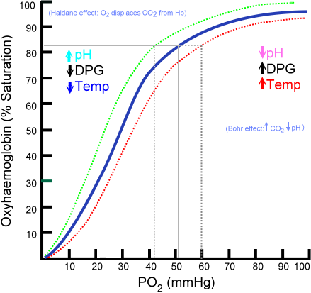
The normal arterial oxygen tension at sea level in healthy people is approximately 85–100 mmHg, depending on age. This equates to an oxygen saturation of 97–100%.
Oxygenation failure is usually defined as an arterial oxygen tension of below 60 mmHg, which equates to an arterial oxygen saturation of 90%.
This level is chosen because, at oxygen tensions below 60 mmHg, oxygen saturation drop quite quickly (due to the sigmoid shape of the oxygen–haemoglobin dissociation curve). Because oxygen delivery to the tissues is a function of cardiac output, haemoglobin and oxygen saturation, oxygen delivery drops quickly at these levels, potentially causing tissue hypoxia and death.

Note: there is very little increase in saturation for increases in PaO2 above 60 mmHg, whereas relatively small changes in PaO2 (partial pressure of oxygen in arterial blood) below 60 lead to large changes in saturation. The PaO2 of normal venous blood is 75%, corresponding to a PaO2 of 40 mmHg.
Ventilatory failure
‘Ventilation’ is the movement of air in and out of the alveoli, and occurs when signals from the brain are transmitted via the spinal cord and nerves to the respiratory muscles (mainly intercostals and diaphram).
The respiratory muscles contract and relax in response, causing expansion and contraction of the chest and the movement of air in and out of the lungs. This mechanism is analogous to a pump whose work depends on the stiffness of the lungs, the resistance in the airways (the energy required to move air through the airways), the stiffness of the chest wall and the body’s metabolic needs. Ventilatory failure occurs either when there is some primary failure in the pump mechanism (brain, spine, nerves or muscle) or the work demanded of the pump is too great.
Carbon dioxide levels are inversely proportional to alveolar ventilation. When ventilatory failure occurs, movement of air in and out of the alveoli is reduced relative to the body’s needs, and consequently, carbon dioxide levels increase. As carbon dioxide levels increase, oxygen tension in the alveoli decreases, leading to arterial hypoxia. Hypoxia may also occur due to an intrinsic lung process that causes ventilatory failure due to excessive work demands, for example, severe pneumonia.
The causes of ventilatory failure can be divided into several broad groups: primary pump failure and excessive work.
1. Primary pump failure
central nervous system (CNS)—stroke, trauma, drugs (morphine, sedatives)
spinal cord—trauma, demyelination, compression, polio
peripheral nerves—demyelination, drugs, trauma, myasthenia gravis
muscles—myositis.
2. Excessive work
parenchymal—pneumonia, cardiac failure, pulmonary fibrosis
airway—asthma, chronic obstructive pulmonary disease (COPD), upper airway obstruction
chest wall—kyphoscoliosis, fractured ribs, pleural thickening/effusions
metabolic—acidosis (diabetic ketoacidosis (DKA), renal failure).
Normal arterial CO2 levels are 35–45 mmHg with an associated pH of 7.35–7.45. Ventilatory failure is said to be present when the arterial pCO2 level is above 45 mmHg.
Treatment
Hypoxia kills, and while supplemental oxygen does not address the underlying problem in ventilatory failure it is vital to prevent tissue hypoxia.
Treatment to reverse alveolar hypoventilation depends on the underlying cause:
- Respiratory depression due to drugs may be reversed with specific antagonists (opiods reversal with naloxone) and hypoventilation due to lung pathology (for example, acute pulmonary oedema (APO) or asthma) may be reversed by specific treatment.
- However, alveolar hypoventilation often requires mechanical assistance with a respiratory pump while the underlying problem is treated. This may be administered non-invasively, via a tight-fitting facemask (Bipap), or invasively, by tracheal intubation. These techniques allow time for treatments to take effect and the underlying process causing hypoventilation to resolve.
Chronic CO2 retainers requiring oxygen therapy often lead to treatment confusion amongst health professionals. As mentioned above, hypoxia kills, and oxygen therapy should not be withheld if indicated. However, this group of patients will require informed health professional management for the duration of that therapy.
Recognising respiratory failure
Because the causes of respiratory failure are varied, some features are common to most cases:
A. Respiratory compensation
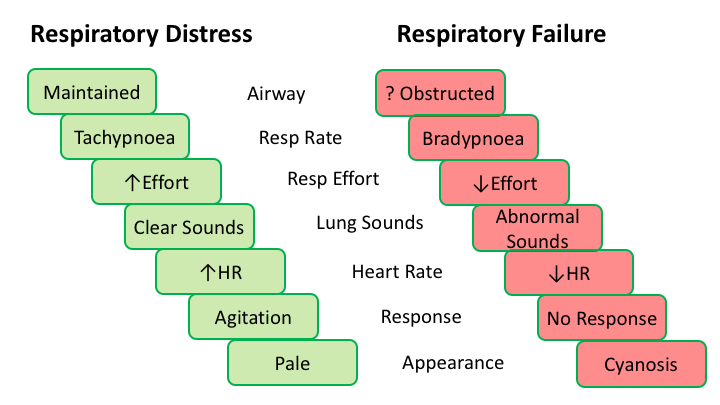
Tachypnoea—tachypnoea is generally present; however, in some causes of ventilatory failure, bradypnoea (low respiratory rate) may be the cardinal sign, for example, narcotic overdose or cerebrovascular accident or terminal event
Use of accessory muscles—in an attempt to increase ventilation or overcome excessive work of breathing, the shoulder girdle, neck and arm muscles are used to augment ventilation
Nasal flaring and head bobbing—a sign of increased respiratory drive
Intercostal, suprasternal or supraclavicular recession—signs of large intrathoracic pressure changes
Paradoxical abdominal movement—the abdomen and chest rise and fall in synchrony in normal respiration; dysynchrony is a sign of impending fatigue and muscle weakness.
- tachycardia
- hypertension
- sweating.
C. End-organ hypoxia
altered mental status:
- hypoxia may cause confusion, agitation or disorientation
- hypercapnia may cause drowsiness or coma
- bradycardia and hypotension (late signs).
D. Haemoglobin desaturation
- cyanosis
- pulse oximetry
- arterial blood gas analysis.


